Interaction Scheme
Molecule
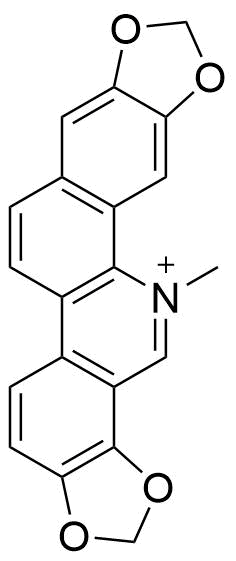
Sanguinarine
c = 0.59 µM
Host
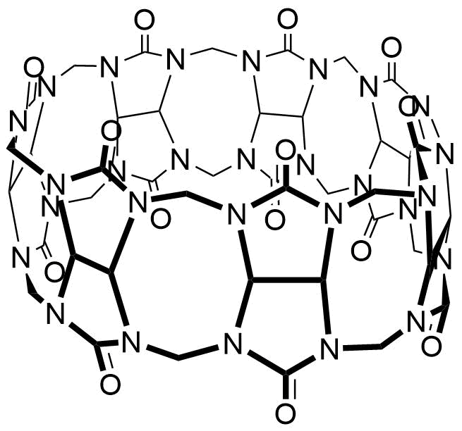
CB7
c = 0.0 — 160.0 µM
Binding Properties
| 𝜈 | Molecule 1 : 1 Host | ||
| Ka = | 1.40⋅106 | ± | M-1 |
| Kd = | |||
| logKa = | |||
| T | 25.0 °C | ||
| Energy | kJ mol-1 | kcal mol-1 | |||
|---|---|---|---|---|---|
| ΔG | = | -35.08 | ± 0.0 | -8.38 | ± 0.0 |
These are the specifications of the determination of the experimental results.
| Detection Method: | Direct | |||
| Assay Type: | Direct Binding Assay | |||
| Technique: | Fluorescence | |||
| 𝛌ex | = | 440.0 nm | ||
| 𝛌em | = | 558.0 nm | ||
| Ibound⁄Ifree | = | 17.7 | ||
Detailed information about the solvation.
| Solvent System | Complex Mixture |
| Solvents | water |
| Additives | hydrochloric acid |
| pH | 4.0 |
Please find here information about the dataset this interaction is part of.
| Citation: |
Z. Miskolczy, L. Biczók, M. Megyesi, G. Tárkányi, R. Mizsei, SupraBank 2026, Inclusion complex formation of sanguinarinealkaloid with cucurbit[7]uril: inhibition of nucleophilic attack and photooxidation (dataset). https://doi.org/10.34804/supra.20210928132 |
| Link: | https://doi.org/10.34804/supra.20210928132 |
| Export: | BibTex | RIS | EndNote |
Please find here information about the scholarly article describing the results derived from that data.
| Citation: |
Z. Miskolczy, M. Megyesi, G. Tárkányi, R. Mizsei, L. Biczók, Org. Biomol. Chem. 2011, 9, 1061–1070. |
| Link: | https://doi.org/10.1039/c0ob00666a |
| Export: | BibTex | RIS | EndNote | |
Binding Isotherm Simulations
The plot depicts the binding isotherm simulation of a 1:1 interaction of Sanguinarine (1.4285714285714285e-05 M) and CB7 (0 — 2.857142857142857e-05 M).
Please sign in: customize the simulation by signing in to the SupraBank.