Interaction Scheme
Molecule
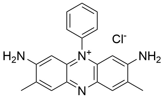
Safranine T
c = 42.0 µM
Host
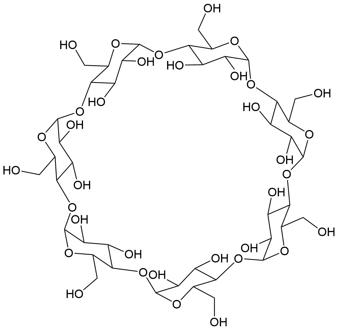
β-CD
Binding Properties
| 𝜈 | Molecule 1 : 1 Host | ||
| Ka = | 160.0 | ± | M-1 |
| Kd = | |||
| logKa = | |||
| T | 20.0 °C | ||
| Energy | kJ mol-1 | kcal mol-1 | |||
|---|---|---|---|---|---|
| ΔG | = | -12.37 | ± 0.0 | -2.96 | ± 0.0 |
These are the specifications of the determination of the experimental results.
| Detection Method: | Direct | |||
| Assay Type: | Direct Binding Assay | |||
| Technique: | Fluorescence | |||
| 𝛌ex | = | 525.0 nm | ||
| 𝛌em | = | 573.0 nm | ||
| Ibound⁄Ifree | = | 2.5 | ||
Detailed information about the solvation.
| Solvent System | Buffer System | 200 mM tris pH-7.2 |
| Solvents | water | |
| Additives | Trometamol | 200.0 mM |
| hydrochloric acid | ||
| Source of Concentration | ||
| Total concentration | 200.0 mM | |
| pH | 7.2 |
Please find here information about the dataset this interaction is part of.
| Citation: |
G. Zhang, S. Shuang, C. Dong, Y. Pang, M. M. Choi, D. Liu, SupraBank 2026, Spectroscopic studies on the interaction of Safranine T with DNA in β-cyclodextrin and carboxymethyl-β-cyclodextrin (dataset). https://doi.org/10.34804/supra.20210928338 |
| Link: | https://doi.org/10.34804/supra.20210928338 |
| Export: | BibTex | RIS | EndNote |
Please find here information about the scholarly article describing the results derived from that data.
| Citation: |
G. Zhang, Y. Pang, S. Shuang, C. Dong, M. M. F. Choi, D. Liu, Journal of Photochemistry and Photobiology A: Chemistry 2005, 169, 153–158. |
| Link: | https://doi.org/10.1016/j.jphotochem.2004.06.016 |
| Export: | BibTex | RIS | EndNote |
Binding Isotherm Simulations
The plot depicts the binding isotherm simulation of a 1:1 interaction of Safranine T (0.125 M) and β-CD (0 — 0.25 M).
Please sign in: customize the simulation by signing in to the SupraBank.