Interaction Scheme
Molecule
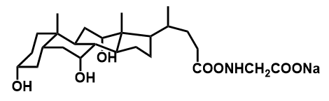
GCA
Host
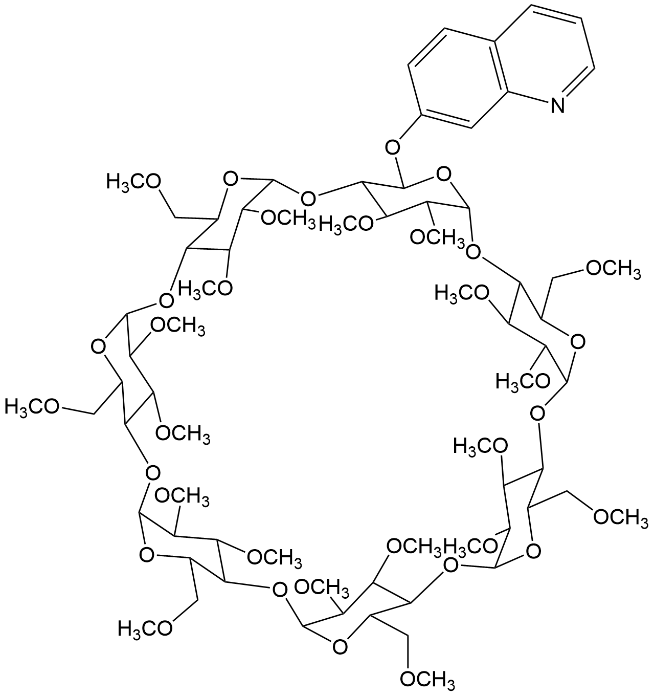
6I-O-(8-hydroxyquinoline)-2I,31- di-O-methylhex...
Binding Properties
| 𝜈 | Molecule 1 : 1 Host | ||
| Ka = | 1.07⋅104 | ± 340.0 | M-1 |
| Kd = | |||
| logKa = | |||
| T | 25.0 °C | ||
| Energy | kJ mol-1 | kcal mol-1 | |||
|---|---|---|---|---|---|
| ΔG | = | -23.0 | ± 0.08 | -5.5 | ± 0.02 |
These are the specifications of the determination of the experimental results.
| Detection Method: | Direct | |||
| Assay Type: | Direct Binding Assay | |||
| Technique: | Fluorescence | |||
| 𝛌ex | = | 300.0 nm | ||
| 𝛌em | = | 398.0 nm | ||
Detailed information about the solvation.
| Solvent System | Buffer System | 100 mM tris pH-7.2 |
| Solvents | water | |
| Additives | Trometamol | |
| hydrochloric acid | ||
| Source of Concentration | ||
| Total concentration | 100.0 mM | |
| pH | 7.2 |
Please find here information about the dataset this interaction is part of.
| Citation: |
D. Guo, Y. Liu, J. Shi, SupraBank 2026, Novel Permethylated β-Cyclodextrin Derivatives Appended with Chromophores as Efficient Fluorescent Sensors for the Molecular Recognition of Bile Salts (dataset). https://doi.org/10.34804/supra.20210928297 |
| Link: | https://doi.org/10.34804/supra.20210928297 |
| Export: | BibTex | RIS | EndNote |
Please find here information about the scholarly article describing the results derived from that data.
| Citation: |
Y. Liu, J. Shi, D.-S. Guo, J. Org. Chem. 2007, 72, 8227–8234. |
| Link: | https://doi.org/10.1021/jo071131m |
| Export: | BibTex | RIS | EndNote | |
Binding Isotherm Simulations
The plot depicts the binding isotherm simulation of a 1:1 interaction of GCA (0.0018709073900841909 M) and 6I-O-(8-hydroxyquinoline)-2I,31- di-O-methylhexakis(2II-VII,3II-VII,6II-VII- tri-O-methyl)-B-cyclodextrin (0 — 0.0037418147801683817 M).
Please sign in: customize the simulation by signing in to the SupraBank.